Path AI at
ASCO 2024
Product Spotlight:
Introducing Path Explore™ IOP and IHC Explore™
Learn more about the products and register for product demonstrations
Path Explore™ PathExplore™ IOP and IHC Explore™ are for research use only. Not for use in diagnostic procedures.

Download the PathAI digital collateral pack for ASCO 2024
Read our posters from ASCO 2024

ASCO 2024 Poster
Association of machine learning (ML)–derived histological features with transcriptomic molecular subtypes in advanced renal cell carcinoma (RCC)
Download
ASCO 2024 Poster
Correlation of immune phenotypes derived from H&E-stained whole slide images with prognosis and response to checkpoint inhibitors in NSCLC.
DownloadAbstract 4519: June 2nd, 2024 - 9:00 AM-12:00 PM
Association of machine learning (ML)–derived histological features with transcriptomic molecular subtypes in advanced renal cell carcinoma (RCC)
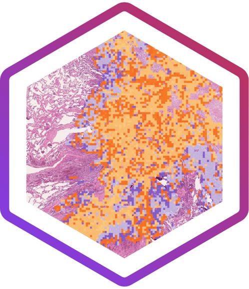
Previously, transcriptomic analysis in the Phase 3 IMmotion 151 (Im151) trial identified 7 molecular subtypes that showed differential outcomes to Atezolizumab+Bevacizumab (A+B) vs Sunitinib (S) treatment. In this abstract, in collaboration with Genentech, human interpretable features (HIFs), including blood vessels, immune cells, fibroblasts, tissue morphologies, and nucleus shape, extracted from H&E-stained whole slide images (WSI) from Im151 and Im150, were used to identify positively associated HIFs within each subgroup in the Im151 WSI and then validated in Im150 molecular subgroups. 169 HIFs were differentially enriched across 3 molecular subsets in both datasets. Our results suggest that clinically relevant RCC subtypes may be extracted directly from H&E-stained WSI and may complement gene expression-based patient stratification and selection strategies.
Abstract 8539: June 3rd, 2024 - 1:30-4:30
Correlation of immune phenotypes derived from H&E-stained whole slide images with prognosis and response to checkpoint inhibitors in NSCLC.
Collaborator: Incendia Therapeutics
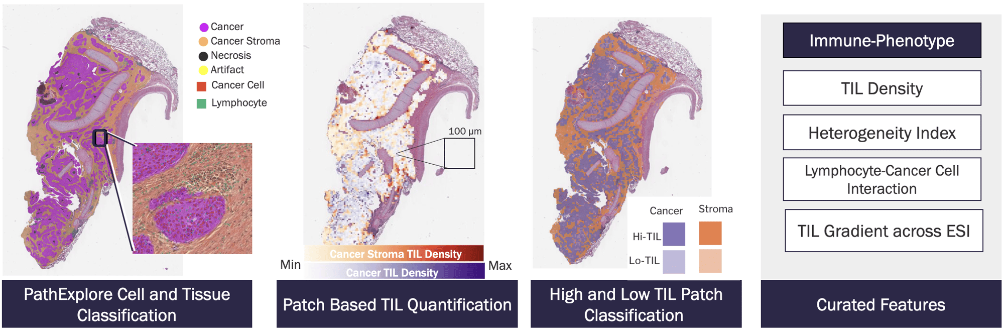
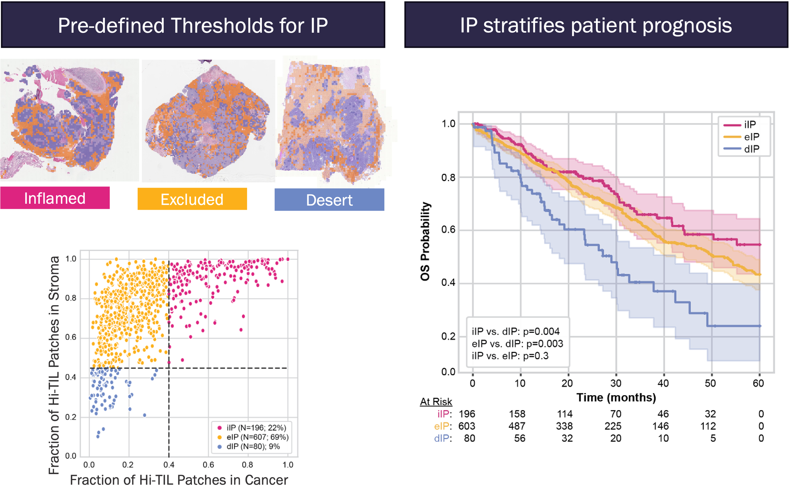
The classification of tumors as inflamed, excluded or desert based on spatial patterns of tumor infiltrating lymphocytes (TILs) is a potential biomarker of patients likely to respond to checkpoint inhibitors (CPI). In this abstract, in collaboration with Incendia Therapeutics, PathExplore IOP is used to classify immune-phenotype (IP) based on patch level TIL distribution in tumor core and periphery from H&E images. Survival analysis indicates Immune inflamed phenotype is associated with improved PFS in CPI-treated NSCLC patients independent of PD-L1 status.
ASCO 2024 Product Spotlight
PathExplore™ IOP
Immuno-Oncology Profiling
Enhance Immune-Oncology Profiling workflows with spatial characterization of tumor-infiltrating lymphocytes (TILs) in the tumor microenvironment (TME) directly from H&E

Identify, quantify, and classify TILs & immune phenotypes with high spatial resolution
- Visualize the density & spatial distribution of TILs with single-cell resolution
- Identify High and Low TIL density regions within tissue regions
- Analyze TIL populations and distributions by tissue compartments
- Quantify cell-cell interactions between tumor and immune cells
- Curated features designed to characterize the tumor-immune microenvironment

PathExplore™ IOP distinct immune profiles (IP) stratifies patient prognosis and treatment response
- Measure % of “High TIL” and “Low TIL” micro patches in each tissue area enables accurate classification of immune phenotypes taking into account heterogeneity of distribution
- Quantitative analytics is used to identify generalizable thresholds for assigning slide-level-based IPs
- Investigations indicate promising applications of IP for patient stratification and novel biomarker discovery

IHC Explore™
AI-Powered Biomarker Quantification
Extract key histopathological features from Immunohistochemistry (IHC) slide images & transform IHC whole-slide images into quantitative insights for next-generation biomarker development

Characterize IHC stains and biomarker expression with spatially-contextualized, single-cell resolution
- Accelerate and enhance the development of AI-enabled biomarkers for novel IHC assays
- Rapid deployment at scale enables continuous quantification of biomarkers
- Deep quantitative measurements of cell types, stain intensity, and completeness
- Identify spatial patterns of stain and expression heterogeneity that are not encapsulated by categorical scores
- Unveil distinct sub-populations that drive novel biomarker discovery and standardization for scoring strategies

CASE STUDY
AI-Assisted Titer Selection in Early Assay Development
- PathAI deployed IHC Explore on prostate cancer specimens stained with a novel, in-development assay
- IHC Explore quantifies staining intensity at single-cell resolution, enabling rapid assay characterization and titer optimization
- Continuous staining intensity measurement provides added value for next-generation biomarkers and precision medicine strategies